My dog has a bump. Can vets diagnose mast cell tumors by microscope?
Updated On September 23rd, 2025
Pet's info: Dog | Mixed Breed | Female | spayed | 9 years and 2 months old | 4 lbs
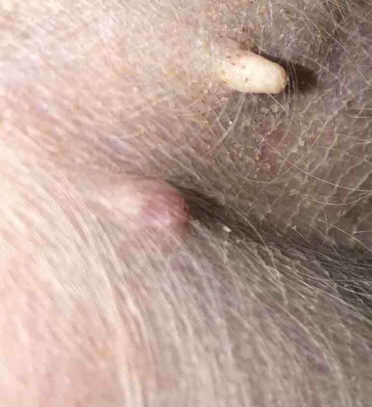
Would a Vet have been able to diagnose a mast cell tumor on a microscope? I don’t want it to be that. Precious got blood tested from a bump & the doctor said she nor anyone else there trained to know what kind it is. She said it was all of the same cell though & I think she said they were tightly packed. She’s having it removed but I’m scared it’s something bad. It has never bothered her. It’s been there since June & has only grown a tiny amount but it’s still small. It got softer afterwards

5 Answers
Most Helpful Answer

Published on April 7th, 2018
Hi, thanks for using Petco Pet Education Center, formerly Petcoach! It depends on the Vet's experience with reading cytology. Most Vets send out their cytology samples to a specialist who is trained to read them. Mast cell tumors have a classic look and tend to be one of the easier tumors to diagnose. You could ask your Vet to send the slides out since that only takes 1-2 days to get them back but sometimes aspirates do not get you an answer. I know you have surgery set up and a biopsy planned. That is the best thing for Precious, to get the mass off and know what it is. It has a high chance of being benign. It is a good thing it hasn't grown much. I wish you the best!
1Pet Parents found this answer helpful
Related Answers from Veterinarians
-
Published on April 7th, 2018
Many veterinarians can identify mast cell tumors under the microscope. However, sometimes they can look different than expected so we can’t say for sure. But if it had all the same cells, it should go. It looks small and your vet should be able to take plenty of surrounding tissue in that area so hopefully that will be the end of it. You can always have that tissue sent to the lab to have it identified for sure. Best of luck to you and Precious!
1Pet Parents found this answer helpful
-
Published on April 7th, 2018
Thank you for submitting your question regarding Precious. A fine needle aspirate is an in office procedure used to diagnose a skin mass. The cells are collected and evaluated under the microscope. A mast cell tumor has distinctive cells and many times can be identified in this way. If the cells are questionable, the slides can be submitted for a review by a pathologist. Alternatively, the mass can be surgically removed and the whole mass submitted to the laboratory for diagnosis. I hope this information helps!
1Pet Parents found this answer helpful
-
Published on April 7th, 2018
Thank you for using Petco Pet Education Center, formerly Petcoach. In many situations surgical removal and histopathology by a trained pathologist is the only way to get a definitive diagnosis. Fine needle aspirate (sticking a needle in mass to get cells) may give an impression of they type of mass, but it is not a firm diagnosis. Many times removal can be curative as well as diagnostic. Best of luck for a good diagnosis and outcome for Precious.
1Pet Parents found this answer helpful
-
Published on December 26th, 2020
Hello, This appears to be a skin growth, not a sore or wound. He is likely licking at it because it is bothering him. This growth looks like one of two very common things, but may be something else entirely as well. I would be suspicious of a benign growth called a Histioctyoma, which is something that generally goes away on its own within a few months without intervention. However, Bandit may need a cone to keep himself from traumatizing it while it runs its course. As well, I am also concerned for something called a mast cell tumor, which is unfortunately a type of cancer that can affect both young and old dogs. Mast cell tumors can have a prognoses range from poor to great, entirely depending on the grade of tumor- this can be determined from a surgical biopsy. I would recommend having Bandit seen by his veterinarian soon. They will likely recommend a test called a fine needle aspirate to look at cytology, or the cells under the microscope. This test can generally tell us if we are dealing with one of the two common masses I just mentioned, and then your vet can guide you on the next best step from there. I hope this helps. Best of luck to you and Bandit.
1Pet Parents found this answer helpful


I took precious to get a bump looked at. The doctor looked at the blood under a microscope & she said it was definitely a mass of some sort. However, she isnt trained to diagnose that type. She said all of the cells were all of the same thing. Prec

Our cat max who is 13 has a lump on his backside we had it checked they stuck it with a needle and said it was a cancerous mast cell tumor and should be surgically removed.Can they actually tell that way and is that the proper procedure.
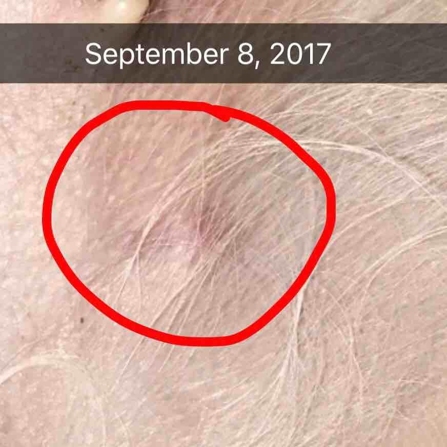
Yesterday she got spayed & also a mass/bump removed. The mass is being sent off next week to be tested. But until then, what could this have been? It has never bothered her nor has she ever bothered it, it was small & only grew a little bit since Jun
Is this round bump as shown in photo is cancerous or what is it exactly and how this can be treated?
Precious has a bump since June on her inner leg. Only grown a little, doesnt bother it & isnt having any other problems. Does this mean its a good chance of being benign? The needle aspiration came back as something the vet couldnt read but it was so
My vet said it's hard to get cells for fine needle aspiration because of the lesion's location. Is that true? The growth suddenly enlarged due to a bacterial infection- I've attached a pic of it from 2 years ago. Is it difficult to get clean margins

The vet tested a tumor on my dogs inner ear and said it has mast cells in it. After removal of ear and testing to see if the cancer has spread she sent the tumor in and it came back it is a histiocytoma tumor. Was the removal of the ear necessary?
I found what looks like a mass on my dogs. Do you think it’s cancer? I’m really worried..
Hi, my 8 month old Staffordshire Bull Terrier has started developing a red lump around 3 weeks ago. It started off as a pea-sized lump when I first noticed it, and has then developed into a significantly wider growth. The first picture attached is a
I took her to the vet and he did an FNA. He said that it might be MCT but he said he wasn't sure. So I figured I would ask. If this is MCT, how serious is this? After searching I learned that FNA isn’t enough to grade MCT but still… can we know anyth

Book an appointment with the pros – our expert vets are here to help.